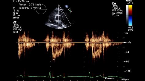
normal 2d echo result|Understanding Normal vs Abnormal Echocardiogram Results : Clark How To Read The 2D Echo Test Result? What Does It Show - Clinico Lottery results for the Florida (FL) Mega Millions and winning numbers for the last 10 draws.
PH0 · What Is 2D EchoTest: Know Uses, Test Results, How To Read It
PH1 · Understanding Normal vs Abnormal Echocardiogram Results
PH2 · Reference (normal) values for echocardiography
PH3 · Interpreting Echocardiogram Results: A Comprehensive Guide for
PH4 · How to read an echocardiogram report
PH5 · How To Read The 2D Echo Test Result? What Does It Show
PH6 · Echocardiography online normal values tables
PH7 · Echocardiography (Normal values)
PH8 · All You Need to Know: How to Read the 2D Echo Test Result
PH9 · A Guide to Understanding Echocardiogram Results
PH10 · 2D Echocardiography Test: Price, Procedure, Uses, Results
Definition of the Tagalog word bangka in English with 6 example sentences, and audio.
normal 2d echo result*******How To Read The 2D Echo Test Result? What Does It Show - Cliniconormal 2d echo resultWhat Is 2D EchoTest: Know Uses, Test Results, How To Read It
All You Need to Know: How to Read the 2D Echo Test ResultHow To Read The 2D Echo Test Result? What Does It Show - ClinicoA normal EF is about 55-65 per cent. It’s important to understand that “normal” is not 100 per cent. Measuring the EF helps your doctor to understand how well the heart is pumping. Generally an EF below 40 per cent is considered a sign that the heart is not pumping as .

A transthoracic echo (TTE), the most common type, involves the patient . Values around 60% (+/- 5% ) are generally considered normal, while values below 30% indicate severe impairment.
Normal (reference) values for echocardiography, for all measurements, according to AHA, ACC and ESC, with calculators, reviews and e-book.normal 2d echo result Understanding Normal vs Abnormal Echocardiogram ResultsThe 2D echo test report involves examining the demographics of the patient, study details, echocardiographic results (chamber dimensions, valve function, and LV function), and . Normal values for M-mode, 2D, Doppler flow, tissue Doppler, speckle strain echocardiography, wall motion, pericardial effusion, exam quality.
A 2D echo test, also known as two-dimensional echocardiography, is a non-invasive procedure that utilises ultrasound waves to evaluate the function and structure of your . The 2D Echo test result includes information like the functioning of the heart, any malfunction, abnormal anatomy or atypical movement of heart structures. Read this .
The resulting image of an echocardiogram can show a big picture image of heart health, function, and strength. For example, the test can show if the heart is enlarged or has thickened walls. Walls thicker .LV and LA can be within the ‘‘normal’’ range for patients with acute severe MR or with chronic severe MR who have small body size, particularly women, or with small LV size preceding the occurrence of MR.
Pumping Ability – Strength of Valves. The doctors evaluate the ejection fraction – EF in order to determine how well the heart is pumping blood. It is a value .
A normal 2D echo test result mentions the absence of any heart malfunction and abnormal anatomy or atypical movement of heart structures. b) What does 60% mean in an echo report? The 60% in a 2D echo test report is the ejection fraction (percentage of blood pumped out of the filled left ventricle with each contraction of the heart). A normal .An echocardiogram (or echo) is an ultrasound of the heart. During an echo, we record short videos of the heart as it beats, and from these videos we can learn about the structure and function of the heart. The left ventricle is the main pumping chamber of your heart – it is the one where blood leaves your heart to be pumped around your body. Normal values for Echocardiography Normal values for Echocardiographic M-mode, 2D, Doppler and Speckle Tracking Strain Measurements and Calculations. January 17th, 2021 (updated May 8th, 2022) Table of Contents. Left ventricular M-mode, 2D, Doppler, tissue Doppler measurements . Tables 1 – 3 (Page 1) Left atrial .
Normal values and thresholds for all heart structures including . (g/m²) - 2D method. 2D methods: truncated ellipsoid method or area length method. 44-88: . Part 1: aortic and pulmonary regurgitation (native valve disease). In European journal of echocardiography : the journal of the Working Group on Echocardiography of the European . Two-dimensional (2D) or three-dimensional (3D) echocardiogram. These images provide pictures of the heart walls and valves and of the large vessels connected to your heart. A standard echocardiogram begins with a 2D study of the heart. A 3D echocardiogram is available in some medical centers and hospitals. It's often done to .Normal or mildly abnormal leaflets: Moderately abnormal leaflets: Severe valve lesions (e.g., flail leaflet, severe retraction, large perforation) Right atrium (Size) Usually normal (RA major 45mm) Normal or mild dilatation: Usually dilated 1 (RA major >45mm) Right ventricle (Size) Usually normal (RVD1 basal 41mm) Normal or mild dilatation .Doppler signals can be colour-coded to enhance visualisation of blood flow (termed Doppler colour-flow mapping) and is the best way to determine the degree of narrowing, calcification or leakage of a valve. 2D Echo/Doppler study is one of the most important non-invasive investigations used in the diagnosis of heart disease today. Echocardiographic reference ranges for normal cardiac chamber size: results from the NORRE study . a variety of mostly dated echocardiographic techniques. 2–4 The Normal Reference Ranges for Echocardiography . diastolic area – end-systolic area)/end-diastolic area. The 2D RV outflow tract diameters were measured from the . Echocardiogram. An echocardiogram . HFpEF can be the result of aging, diabetes, or high blood pressure. . A normal ejection fraction range is between 52 and 72 percent for men and between 54 . A normal test result reflects normal functioning; structure; and movement of heart muscles, valves and chambers. An abnormal 2D ECHO (both TTE and TEE) test could indicate a myriad of heart problems.It requires consultation with a cardiologist for a final diagnosis and treatment. Two-dimensional (2D) ultrasound. This approach is used most often. . For example, some people with valve disease need echo tests on a regular basis. You’re preparing for a surgery or procedure. Your provider wants to check the outcome of a surgery or procedure. . How do I get the results of my test?

Patients with uncomplicated viral pericarditis have normal echocardiogram results. 1. Aorta. The diameter of the aortic root is measured routinely. Sometimes it is possible to identify dilation of the ascending aorta, the arch, or the descending aorta. . Principles and practice of echocardiography. 2nd ed. Philadelphia, Pa: Lea and .Understanding Normal vs Abnormal Echocardiogram Results Patients with uncomplicated viral pericarditis have normal echocardiogram results. 1. Aorta. The diameter of the aortic root is measured routinely. Sometimes it is possible to identify dilation of the ascending aorta, the arch, or the descending aorta. . Principles and practice of echocardiography. 2nd ed. Philadelphia, Pa: Lea and . Paediatric echo reports tend to follow a standard format as defined by guidelines published by the American Society of Echocardiography. 2 This includes minimum elements, such as the patient's characteristics, clinical data, echo findings (i.e. defining structural anatomy, measurements of cardiovascular structures, haemodynamic .2D echo assessment. SCCS Zabrze How do we see the aortic valve ? Planar views of valve anatomy in standard views. . regurgitation with anatomically normal aortic valve and ascending aorta aneurysm by TEE European Journal of Echocardiography (2010) 11, 645–658. SCCS Zabrze Aortic valve repairability Recommendations of echocardiography in the current hypertension guidelines. In the 2013 ESH/ESC Guidelines for the management of arterial hypertension, echocardiography is the second-line study based on medical history, physical examination, and findings from routine laboratory tests [].The guidelines recommended .
The following are three learning modules are designed to provide a basic introduction in interpretive echocardiography for the following anatomy: Normal Anatomy Atrial Septal Defects (ASDs)3-D echo technique captures three-dimensional views of the heart structures with greater detail than 2-D echo. The live or "real time" images allow for a more accurate assessment of heart function by using measurements taken while the heart is beating. 3-D echo shows enhanced views of the heart's anatomy and can be used to determine the .Normal reference ranges depend on sex, body surface area (except in extreme obesity) and age (often useful to use an indexed value or a Z-score for children / adolescents): Normal male ascending aorta size = 30 mm ± 4 mm (indexed is up to 1.5 ± 2 mm/m 2) Normal female ascending aorta = 27 mm ± 4 mm (indexed is up to 1.6 ± 3 mm/m 2)
Filipino Sex Stories Init, Sarap, At Pantasya(ang Unang Yugto) Last Part: Repost/re-edit By Xelrekusuta Para akong estatwa nang marinig ko ang response ni chie sa tanong ko kung masarap ba ang tagpo nila ni Maris. Nag iinit ako sa oras na .
normal 2d echo result|Understanding Normal vs Abnormal Echocardiogram Results