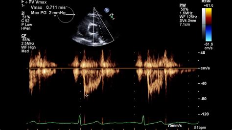
2d echo normal results|“If My Echo Is Normal, Is My Heart OK?’ : Manila Normal 2D measurements: LV minor axis ≤ 2.8 cm/m 2, LV end-diastolic volume ≤ 82 ml/m 2, maximal LA antero-posterior diameter ≤ 2.8 cm/m 2, maximal LA volume ≤ 36 ml/m 2 (2;33;35). ∗∗ . 5J646 Flight Tracker - Track the real-time flight status of Cebu Pacific 5J 646 live using the FlightStats Global Flight Tracker. See if your flight has been delayed or cancelled and track the live position on a map.
PH0 · “If My Echo Is Normal, Is My Heart OK?’
PH1 · What is a 2D Echo Test?
PH2 · What is 2d Echo test and its Uses, Test Results, and Normal
PH3 · Reference (normal) values for echocardiography
PH4 · Normal Echo Values
PH5 · Interpreting Echocardiogram Results: A Comprehensive Guide For
PH6 · Interpreting Echocardiogram Results: A Comprehensive Guide
PH7 · Echocardiography online normal values tables
PH8 · Echocardiography (Normal values)
PH9 · Echocardiographic reference ranges for normal cardiac
PH10 · Echocardiogram: What It Shows, Purpose, Types, and Results
PH11 · Echocardiogram: What It Shows, Purpose, Types, and Results
PH12 · Echocardiogram
PH13 · Echocardia
Before TuneCore, artists needed a label to get their music sold online. In 2006, we disrupted the industry by partnering directly with Digital Stores to allow any musician to sell their songs worldwide. Today, TuneCore is the world's leading digital music aggregator. Choose an unlimited distribution plan, upload your music, and we'll do the rest.
2d echo normal results*******Normal (reference) values for echocardiography, for all measurements, according to AHA, ACC and ESC, with calculators, reviews and e-book.TTE Echo normal reference values (Tables, charts). Data collected from 10 pdf guidelines in one place.
Two-dimensional (2D) or three-dimensional (3D) echocardiogram. A 2D echo is the standard test, which shows your doctor images of your heart's walls, valves, . In this guide, we will explore the different types of echocardiography, delve into the components of an echocardiogram report, discuss the interpretation of normal .2d echo normal results In this guide, we will explore the different types of echocardiography, delve into the components of an echocardiogram report, discuss the interpretation of normal . Normal 2D measurements: LV minor axis ≤ 2.8 cm/m 2, LV end-diastolic volume ≤ 82 ml/m 2, maximal LA antero-posterior diameter ≤ 2.8 cm/m 2, maximal LA volume ≤ 36 ml/m 2 (2;33;35). ∗∗ .

Normal values and thresholds for all heart structures including illustrations where to measure the relevant values.
Normal values for M-mode, 2D, Doppler flow, tissue Doppler, speckle strain echocardiography, wall motion, pericardial effusion, exam quality. Recently, the 2D sub-study of the NORRE Study has been published providing normal 2D-echocardiographic reference values for left and right heart .
Two-dimensional (2D) or three-dimensional (3D) echocardiogram. These images provide pictures of the heart walls and valves and of the large vessels connected to your heart. A .
A two-dimensional Echocardiogram or 2D Echo test is a diagnostic test that uses ultrasound waves to assess the functioning of the heart. When these waves hit the organ . Visit my website: https://www.discoverecho.comAn 82 year old female needed a complete 2D echocardiogram with Doppler for Sarcoid. With sarcoid, the valves, .Typically, there is no required recovery time after an echocardiogram. You can resume your normal activities immediately after the test. Depending on the results, the doctor may provide further recommendations. . What do the Results of a 2D Echo Test Show? A 2D Echo test provides valuable information about the heart, including: 1. Changes in .Get complete information on the 2d Echo test: Procedure, uses, results interpretation, and normal range. Understand how this test is used to diagnose. 24/7 Appointment Helpline +91 40 4567 4567. International +91 40 6600 0066. Home. About. . 2D Echo test is a non-invasive diagnostic test used for detecting various cardiac abnormalities like.Eccentric hypertrophy. >115 (Male) >95 (Female) <0,42. Concentric remodeling. ≤115 (Male) ≤95 (Female) >0,42. Description of LV geometry, using at the minimum the four categories of normal geometry, concentric remodelling, and concentric and eccentric hypertrophy, should be a standard component of the echocardiography report.Two-dimensional (2D) ultrasound is the most commonly used modality in echocardiography. The two dimensions presented are width (x axis) and depth (y axis). The standard ultrasound transducer for 2D echocardiography is the phased array transducer, which creates a sector shaped ultrasound field (Figur 1).Echocardiogram. An echocardiogram is a noninvasive (the skin is not pierced) procedure used to assess the heart's function and structures. During the procedure, a transducer (like a microphone) sends out sound waves at a frequency too high to be heard. When the transducer is placed on the chest at certain locations and angles, the sound waves .
The NORRE (Normal Reference Ranges for Echocardiography) study is the first European, large, prospective, multicentre study performed in 22 laboratories accredited by the European Association of Cardiovascular Imaging (EACVI) and in 1 American laboratory, which has provided reference values for all 2D echocardiographic .
2D Echocardiography/Doppler Study (Trans thoracic 2D Echo/Doppler Study) This is a non-invasive, painless and risk-free heart scan using high frequency ultrasound waves reflecting off various structures of the heart to obtain real-time images (in one and two dimensions) of your beating heart. When combined with the Doppler technique, which .
An echocardiogram (or echo) is an ultrasound of the heart. During an echo, we record short videos of the heart as it beats, and from these videos we can learn about the structure and function of the heart. The left ventricle is the main pumping chamber of your heart – it is the one where blood leaves your heart to be pumped around your body.
Usually, an EF falls between 50% and 70%. Valve Function: The 2D Echo assesses the function of heart valves, including the mitral, aortic, tricuspid, and pulmonary valves. The report will note whether the valves are functioning normally or if there is regurgitation (leakage) or stenosis (narrowing). Chamber Sizes: The measurements of the heart .The results of a 2D echo test are available after the test. A cardiologist interprets these results and can include the following parameters: . The 2D echo test results are considered normal if there are no signs of structural abnormalities or functional issues. If the heart shows abnormalities, it may indicate conditions that require further . An echocardiogram can detect many different types of heart disease. These include: Congenital heart disease, which you’re born with. Cardiomyopathy, which affects your heart muscle. Infective endocarditis, which is an infection in your heart’s chambers or valves. Pericardial disease, which affects the two-layered sac that covers the outer .
A normal test result reflects normal functioning; structure; and movement of heart muscles, valves and chambers. An abnormal 2D ECHO (both TTE and TEE) test could indicate a myriad of heart problems. It requires consultation with a cardiologist for a final diagnosis and treatment. A normal stress echocardiogram reveals that the heart .
Palpitations or increased heart beat are feelings or sensations that your heart is pounding or racing. They can be felt in your chest, throat, or neck. You may Have an unpleasant awareness of your own heartbeat or may Feel like your heart skipped or stopped beats The heart's rhythm may be normal or abnormal when you have palpitations. Normal values for Echocardiography Normal values for Echocardiographic M-mode, 2D, Doppler and Speckle Tracking Strain Measurements and Calculations. January 17th, 2021 (updated May 8th, 2022) Table of Contents. Left ventricular M-mode, 2D, Doppler, tissue Doppler measurements . Tables 1 – 3 (Page 1) Left atrial .

Taken at an interval, this test can also show the improvement or deterioration of patient’s angina. A negative TMT or Stress Test is declared when the patient can reach a certain heart rate without showing any ECG changes. This rate is known as target heart rate and it is also calculated by a formula (Target Heart Rate = 220 – age of patient).
2d echo normal results “If My Echo Is Normal, Is My Heart OK?’ An echo test can allow your health care team to look at your heart’s structure and check how well your heart functions. The test helps your health care team find out: The size and shape of your heart, and the size, thickness and movement of your heart’s walls. How your heart moves during heartbeats. The heart’s pumping strength.“If My Echo Is Normal, Is My Heart OK?’ Transthoracic echocardiography (TTE), sometimes called “surface echocardiography,” is a basic tool for investigation and follow-up of heart disease. Consultants who interpret TTE endeavour to provide accurate, useful reports to colleagues who order these tests. Referring physicians sometimes find reported results difficult to .
Contact Numbers and Email Address. Phone: (505) 476-4622 Fax: (505) 476-4620 Email: [email protected] Help Desk: [email protected] Staff Directory: Ruth Romero – Senior Board Administrator Roxann Ortiz-Peña – Senior Health Licensing & Support Specialist
2d echo normal results|“If My Echo Is Normal, Is My Heart OK?’