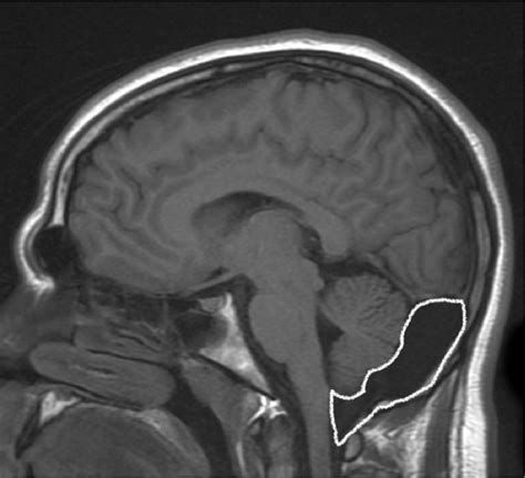
mega cisterna magna versus arachnoid cyst|Mega cisterna magna : Bacolod Mega cisterna magna needs to be distinguished from other causes of an enlarged retrocerebellar CSF space: 1. arachnoid cyst: can be difficult to distinguish . Tingnan ang higit pa Get the free Chess.com app for your device and play chess games with friends around the world! Solve puzzles, take lessons, play vs. computers & more!Total War: WARHAMMER III. All Discussions Screenshots Artwork Broadcasts Videos Workshop News Guides Reviews . If you left click and select a spell, you can right click to get off the selected spell. If you have selected a spell and accidentally started casting, if the spell is out of range and the character is moving within range, you .
PH0 · Mega cisterna magna, arachnoid cyst, and Dandy Walker variant
PH1 · Mega cisterna magna
PH2 · Mega Cisterna Magna and Retrocerebellar Arachnoid Cysts
PH3 · Mega Cisterna Magna Versus Arachnoid Cyst
PH4 · Mega Cisterna Magna
PH5 · Is it an arachnoid cyst or a mega cisterna magna? What to and
PH6 · CSF flow study in differentiation between the arachnoid cyst and mega
PH7 · CSF flow study in differentiation between the
PH8 · Arachnoid cyst
PH9 · Arachnoid Cysts: What Are They, Location, Causes & Symptoms
PH10 · Arachnoid Cyst Imaging
Suggested browser Microsoft Edge, Chrome or Safari only. Total Hits : 511,854,938 | Yearly Hits : 15,903,579 | Monthly Hits : 43,063,989
mega cisterna magna versus arachnoid cyst*******A mega cisterna magna is thought to occur in ~1% of all brains imaged postnatally. It constitutes 54% of all cystic posterior fossa malformations 4. Especially if noted antenatally, a mega cisterna magna has been associated with: 1. infarction 2. inflammation/infection: particularly cytomegalovirus 3. . Tingnan ang higit pa
Some authors have proposed that mega cisterna magna is a result of a delayed Blake pouch fenestration; when fenestration . Tingnan ang higit pa
On antenatal ultrasound, mega cisterna magna refers to an enlarged retrocerebellar CSF space: 1. usually >10 mm (some . Tingnan ang higit paMega cisterna magna needs to be distinguished from other causes of an enlarged retrocerebellar CSF space: 1. arachnoid cyst: can be difficult to distinguish . Tingnan ang higit pamega cisterna magna versus arachnoid cystThe term was coined by the Belgian neurosurgeon Richard Gonsette (1929-2014)8in 1962, in patients with cerebellar atrophy 7. Tingnan ang higit paMay 29, 2023 — Mega cisterna magna refers to a cystic posterior fossa malformation that is characterized by an enlarged cisterna magna, absence of hydrocephalus, and an intact cerebellar vermis. It must be .
Okt 14, 2019 — Mega cisterna magna, arachnoid cyst, and Dandy Walker variant. Midline posterior fossa fluid collections in adults usually represent benign congenital enlargement of the the cisterna magna.
Ene 10, 2017 — There is debate as to whether mega cisterna magna (MCM) arises from a pathologic insult; recent evidence suggests that it may be on the mildest end of the .
Mega Cisterna Magna and Arachnoid are both types of cysts that occur in the central nervous system and contain cerebrospinal fluid, or CSF. While they are very similar, it is important to make a correct diagnosis between .
Okt 9, 2021 — Arachnoid cysts usually grow on the brain (intracranial arachnoid cysts). Less commonly, they grow on the spinal cord (spinal arachnoid cysts). Both types .Mar 12, 2021 — Most arise as developmental anomalies. A small number of arachnoid cysts are associated with neoplasms. CT imaging is often sufficient to make the diagnosis, but when additional information is.

Abr 24, 2020 — The main challenge in the diagnosis of posterior fossa cysts in the paediatric age group is differentiating between arachnoid cysts from mega cisterna magna. Mega cisterna magna is the enlarged cisterna .Mega cisterna magna Abr 24, 2020 — The main challenge in the diagnosis of posterior fossa cysts in the paediatric age group is differentiating between arachnoid cysts from mega cisterna magna. Mega cisterna magna is the enlarged cisterna .Hun 11, 2024 — Arachnoid cysts are relatively common benign and asymptomatic lesions occurring in association with the central nervous system, both within the intracranial compartment (most common) as well .Is it an arachnoid cyst or a mega cisterna magna? What to and where to look for to make the correct diagnosis? Congress: ECR 2018. Poster Number: C-1854. Type: Educational .Hun 11, 2024 — Arachnoid cysts are relatively common benign and asymptomatic lesions occurring in association with the central nervous system, both within the intracranial compartment (most common) as well .
Another well-known common finding of arachnoid cysts is scalloping of the adjacent bone, in this case occipital bone, due to gradual increase in size of the cyst in the development stage, thus applying a mass efect on bone, .Abr 24, 2020 — Mega cisterna magna is the enlarged cisterna magna (more than 10 mm in mid-sagittal plane) with intact cerebellar vermis and normal fourth ventricle. It freely communicates with the subarachnoid .Mega Cisterna Magna and Arachnoid cysts look similar on MRI read outs so in order to correctly diagnosis a ventriculogram should be performed with the injection of dye into the blood or with a catheter into the heart. The Mega cisterna magna will show communication with the subarachnoid space indicating a normal cerebellar vermis and fourth .The management of posterior fossa arachnoid cyst (PFAC) in adults is controversial. To review our cases and literature, propose a practically useful surgical strategy, which gives excellent long-term outcome in management of PFAC. . which they had reported and that included mega cisterna magna (11 cases), Dandy-Walker malformation (5 cases .What are mega cisterna magna (MCM) and arachnoid cysts (AC)? MCM involves the enlargement of normal fluid-filled space in the brain with no other structural differences anomaly in other cerebral structures. ACs are usually benign, cerebro-spinal fluid-like collections that develop within the layers of the membranes that wrap the brain. .Ene 15, 2015 — Posterior fossa cysts and cyst-like malformations (Blake’s pouch cyst, arachnoid cysts, and mega cisterna magna). In: Boltshauser E , Schmahmann JD , eds. Cerebellar disorders in children . London, England: Mac Keith, 2012 ; 212–216.
Mar 12, 2021 — A large cisterna magna (mega cisterna magna) occasionally may be confused with an arachnoid cyst. Mega cisterna magna may represent a normal variant (intact cerebellum and vermis), but it may be associated with Dandy-Walker syndrome, either full blown or a variant in which the vermis is either completely or partially absent.How do MCM and AC cysts happen? Both are considered cystic lesions of the posterior brain. In the early development of the fetus an alteration occurs in the making of the system and spaces where the spinal fluid normally travels, and mega cisterna magna results as a consequence of the accumulation of spinal fluid in this space.Mega cisterna magna refers simply to focal enlargement of the cisterna magna, the biggest of the subarachnoid cisterns, which is located posterior and inferior to cerebellum and dorsal surface of the brain stem, medulla oblongata, and superior to foramen magnum. [1] The most accepted quantitative measurement of the enlargement is more than 10 .Abr 22, 2024 — infravermian cyst that communicates with fourth ventricle. cyst is smooth with thin walls that can be visualized on thin sagittal T2 images. it can impress on medial side of cerebellar tonsils due to size .Hun 28, 2020 — 18 Mega Cisterna Magna Cole T. Lewis, Octavio Arevalo, Rajan P. Patel, and David I. Sandberg. 18.1 Case Presentation. 18.1.1 History. A 15-year-old female patient presents with a history of a one .Although differential of retrocerebellar arachnoid cysts and mega cisterna magna is of little clinical concern, as both lesions are mostly asymptomatic and require no follow-up, they are both frequently encountered lesions in .May 29, 2023 — Similar to mega cisterna magna, arachnoid cysts match the CSF signal density on computed tomography (CT) and signal intensity on magnetic resonance imaging (MRI) and typically do not enhance following administration of intravenous contrast. In contrast to mega cisterna magna, arachnoid cysts may be associated with a mass .
Ene 15, 2015 — Posterior fossa cysts and cyst-like malformations (Blake’s pouch cyst, arachnoid cysts, and mega cisterna magna). In: Boltshauser E , Schmahmann JD , eds. Cerebellar disorders in children . London, England: Mac Keith, 2012 ; 212–216.

Mar 26, 2023 — Arachnoid cysts (ACs) are usually congenital, extraaxial, benign, cystic lesions filled with cerebrospinal fluid. The most common etiology is splitting of the arachnoid layer. . In mega cisterna magna, cystic enlargement of the subarachnoid space, connected to the fourth ventricle by the foramen of Magendie, is observed [30, .
mega cisterna magna versus arachnoid cyst Mega cisterna magna Mar 26, 2023 — Arachnoid cysts (ACs) are usually congenital, extraaxial, benign, cystic lesions filled with cerebrospinal fluid. The most common etiology is splitting of the arachnoid layer. . In mega cisterna magna, cystic enlargement of the subarachnoid space, connected to the fourth ventricle by the foramen of Magendie, is observed [30, .Cystic or cyst-like malformations of the posterior fossa represent a spectrum of disorders, including the Dandy-Walker malformation, vermian-cerebellar hypoplasia, mega cisterna magna, and arachnoid cyst. Differentiation of these lesions may be difficult with routine cross-sectional imaging; however .Abr 22, 2024 — infravermian cyst that communicates with fourth ventricle. cyst is smooth with thin walls that can be visualized on thin sagittal T2 images. it can impress on medial side of cerebellar tonsils due to size cyst does not communicate with the cisterna magna posteriorly. upward displacement of the vermis. no vermian hypoplasia or rotation
RotoWire breaks down the top players to consider on the waiver wire heading into Week 16 of the 2023-24 NBA season.
mega cisterna magna versus arachnoid cyst|Mega cisterna magna