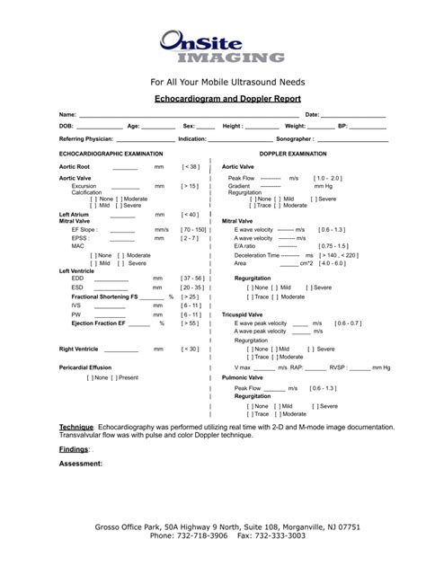
normal echocardiography report|echocardiography report sample : Pilipinas Find the normal ranges and abnormal thresholds for various echocardiographic parameters, such as left and right ventricle size, mass, volume, function, pressure, and kinetics. Compare the values . Laguna: City: Santa Rosa: Postal code: 4026: Population (2020) 39,496: Philippine major island(s) Luzon: Coordinates: 14.2902, 121.1095 (14° 17' North, 121° 7' East) Estimated elevation above sea level: 17.6 meters (57.7 feet) Dila is a barangay in the city of Santa Rosa, in the province of Laguna. Its population as determined by the 2020 .
PH0 · standard range for echocardiogram results
PH1 · normal values on echocardiogram
PH2 · normal ranges for echocardiogram results
PH3 · normal echo report format
PH4 · how to read echocardiography report
PH5 · echocardiography report sample
PH6 · echocardiography normal values
PH7 · ase normal values for echocardiogram
PH8 · Iba pa
This web site contains age-restricted materials. You must be 18 or older to access this website. This website contains age-restricted materials.
normal echocardiography report*******Find normal values for aorta, atria, ventricles, valves and pulmonary veins in 2D echocardiography. Learn how to assess valvular regurgitation and stenosis with Doppler parameters and qualitative criteria.Find the normal ranges and abnormal thresholds for various echocardiographic parameters, such as left and right ventricle size, mass, volume, function, pressure, and kinetics. Compare the values .

A normal EF is about 55-65 per cent. It’s important to understand that “normal” is not 100 per cent. Measuring the EF helps your doctor to understand how well the heart is .Normal values and thresholds for all heart structures including illustrations where to measure the relevant values. Below is a complete and thorough list of normal echo values. This list of normal echo values is from echopedia.org. Left Ventricle. Left Ventricular Systolic Function. Left Ventricular Diastolic . The echocardiogram report evaluates the size and function of the cardiac chambers, including the left ventricle, right ventricle, left atrium, and right atrium. .
Echo is the cheapest and least invasive method available for screening cardiac anatomy. Generalists most commonly request an echo to assess left ventricular (LV) dysfunction, to rule out the heart as a . Modern echocardiography either reports diastolic function as normal or grades DD by class (1 through 4). 13 Class 1 DD (impaired myocardial relaxation) was . Abstract. The echocardiogram is a readily accessible imaging modality for GPs investigating patients with suspected cardiac structural abnormalities. The most .
The adult transthoracic echocardiography report should be comprised of the following sections: 1) Demographic and other Identifying Information, 2) Echocardiographic .
An echocardiogram uses sound waves to create pictures of the heart. This common test can show blood flow through the heart and heart valves. Your health care provider can use the pictures from the test to find heart disease and other heart conditions. Other names for this test are: Heart ultrasound. Heart sonogram. 1.5 ± 0.5 (0.5-2.5) 0.9 ± 0.4 (0.1-1.7) Data are expressed as mean ± SD (95% confidence interval). Note that for e´ velocity in subjects aged 16 to 20 years, values overlap with those for subjects aged 21 to .
normal echocardiography report An echocardiogram is a specialized ultrasound scan of the heart. It gives detailed information about how efficiently the heart pumps blood and oxygen to the organs and how well the heart valves work. A trained and qualified cardiologist (heart specialist) needs to read an echocardiogram to know if the results are normal or not.
An echo test can allow your health care team to look at your heart’s structure and check how well your heart functions. The test helps your health care team find out: The size and shape of your heart, and the size, thickness and movement of your heart’s walls. How your heart moves during heartbeats. The heart’s pumping strength.The following are three learning modules are designed to provide a basic introduction in interpretive echocardiography for the following anatomy: Normal Anatomy. Atrial Septal Defects (ASDs) Ventricular Septal Defects (VSDs) Each learning module provides an overview which outlines the basic echocardiographic views for each lesion including all .

When reporting echo features in VHD it is very important to provide information on the type and degree of valve dysfunction as well as on the hemodynamic burden induced by the valve defect to optimize diagnosis, stratify prognosis, and address management. . The report shall include normal reference values to differentiate .echocardiography report sample When reporting echo features in VHD it is very important to provide information on the type and degree of valve dysfunction as well as on the hemodynamic burden induced by the valve defect to optimize diagnosis, stratify prognosis, and address management. . The report shall include normal reference values to differentiate . In this study, we report normal reference ranges for cardiac chambers size obtained in a large group of healthy volunteers accounting for gender and age. Echocardiographic data were acquired using state-of-the-art cardiac ultrasound equipment following chamber quantitation protocols approved by the European Association of .
1. Ask your doctor how big your heart is. If your heart is enlarged or the walls of your heart have become thicker, this can be in indication of several problems. For example, the doctor will likely measure the thickness of the wall of the left ventricle (the major pumping chamber of the heart).Pre-programmed formulae to automatically calculate all of the commonly required measurements for a formal study. Summary boxes next to each category to allow reporting adjacent to the measured values. Overall summary page to finish your report. Customisable header in the critical care echo template to input whichever images/titles . An echocardiogram, on the other hand, shows a detailed view of the heart's internal structure and how blood flows through it. The test: Measures the size and shape of your heart. Shows how well .Normal values. Echo protocol, summary, report for the patient. Online free echocardiography learning platform and examination software. All measurements (Images and videos). Normal values. Echo protocol, summary, report for the patient. Echocardiography: EN / SK. TECH mED.sk. New examination. Left ventricle. Roughly speaking, echocardiogram uses ultrasound waves to . visualise cardiac structures – in 2D or 3D. measure flow velocities via Doppler. While there may be different echocardiogram report formatting, the interrogated structures are either cardiac chambers or cardiac valves.Echo Report Summary and Conclusions Templates. Normal (Young patient) Normal left ventricular size and systolic function (EF 60 %). Normal diastolic function. Normal (Elderly patient) CAD with RWMA: Normal left ventricular size with preserved systolic function (EF 60 %). Regional wall motion abnormalities are suggestive of coronary artery disease.Clinical echocardiography made easy. Clinical Echocardiography enables you to use echocardiography to its fullest potential in your initial diagnosis, decision making, and clinical management of patients with a wide range of heart diseases. This e-book helps you master what you need to know to obtain the detailed anatomic and physiologic . An echocardiogram can detect many different types of heart disease. These include: Congenital heart disease, which you’re born with. Cardiomyopathy, which affects your heart muscle. Infective endocarditis, which is an infection in your heart’s chambers or valves. Pericardial disease, which affects the two-layered sac that covers the outer . In this NL I will provide you with exactly such a template for echocardiography, together with an explanation. The chambers of the heart All structures that were examined are also mentioned. Important measurements are included in the text. Even though this is a normal exam, it is still important to include all the components that .Two-dimensional (2D) ultrasound is the most commonly used modality in echocardiography. The two dimensions presented are width (x axis) and depth (y axis). The standard ultrasound transducer for 2D echocardiography is the phased array transducer, which creates a sector shaped ultrasound field (Figur 1).
Points are more than a valuable feature, especially if you plan to create DeviantArt Commissions – you can only do that with the points. 4) Sell Fine Art Prints Through The DeviantArt Print Program. I shouldn’t keep covering how to sell art on DeviantArt without taking the time to mention the site’s print program:
normal echocardiography report|echocardiography report sample