scanning electron microscopy slideshare|Scanning electron microscopy : Manila SCANNING ELECTRON MICROSCOPE (SEM) A scanning electron microscope (SEM) is a type of electron microscope that images a sample by scanning it . 300 Welcome Spins on Wild Wild Safari . Using the SAVANNA code on your first 4 deposits for all games except the titles by Dragon provider, All Roulette Games, Ride 'em Poker, Baccarat, Pai Gow, Craps, Caribbean Poker, Top Card Trumps (Casino War), Draw High Low, Pontoon 21, Pirate 21, Red Dog, Oasis Poker. The minimum deposit is $30; .
PH0 · scanning electron microscope (SEM)
PH1 · Scanning electron microscopy (sem)
PH2 · Scanning electron microscopy
PH3 · Scanning electron microscope
PH4 · Scanning Electron microscopy
PH5 · Scanning Electron Microscopy (SEM) lecture
PH6 · Scanning Electron Microscope (SEM)
Sachzna bravely shares her experiences of childhood traumas and heartbreaks, and how she healed from them.
scanning electron microscopy slideshare*******SCANNING ELECTRON MICROSCOPE (SEM) A scanning electron microscope (SEM) is a type of electron microscope that images a sample by scanning it .scanning electron microscope (SEM) Feb 26, 2018 •. 133 likes • 130,424 views. .SuganyaPaulraj. The scanning electron microscope (SEM) was first developed .
Scanning electron microscopy (sem) SEM is a type of electron microscope .
scanning electron microscope (SEM) Feb 26, 2018 •. 133 likes • 130,424 views. Himanshu Dixit. A scanning electron microscope (SEM) is a type of electron . SEM is a type of electron microscope that produces images of a sample by scanning it with a focused beam of electrons in a raster scan pattern. The electrons .
Aug 29, 2016 •. 236 likes • 88,123 views. Saurabh Bhargava. Follow. a brief lecture on SEM; principles, working, instrumentation, and its forensic applications. Education. 1 of 52. Scanning Electron Microscopy (SEM) . SuganyaPaulraj. The scanning electron microscope (SEM) was first developed in 1937 and improved upon in later decades. It uses a beam of electrons to scan sample surfaces at high magnification . Scanning electron microscopy In scanning electron microscopy (SEM) an electron beam is focused into a small probe scan the sample in a raster scan pattern. Several interactions with the sample that .
Scanning electron microscopy (sem) SEM is a type of electron microscope designed for directly studying the surfaces of solid objects, that utilizes a beam of . DEFINITION Scanning electron microscope is a type of electron microscope that images a sample by scanning it with a beam of electrons in a raster scan pattern. WORKING PRINCIPLE In SEM, an .

S. Shivaji Burungale Follow. Dr. S. H. Burungale's document discusses scanning electron microscopy. It notes that Ernst Ruska won the Nobel Prize in . Ganesh Shinde Follow. It is an Microscopic technique that uses an electron as a source for visualizing microscopic parts and has a more resolution than optical microscope. Health & Medicine. 1 of 42. .
6. Principle of SEM • A scanning electron microscope (SEM) is a type of electron microscope that produces images of a sample by scanning it with a focused beam of electrons. • The electrons interact . 6. Basic types of Electron microscopy TEM – The Transmission Electron Microscope was the first type of Electron microscope to be developed and is patterned exactly on the light .
Electron microscope ppt - Download as a PDF or view online for free. . namely Transmission Electron Microscope (TEM) – allows one the study of the inner surface. Scanning Electron Microscope (SEM) – .
Scanning Electron Microscope a Totally Different Imaging Concept • High energy electron beam is used to excite the specimen and the signals are collected and analyzed so that an image can be constructed. • The signals carry topological, chemical and crystallographic information, respectively, of the samples surface. 16 likes • 6,373 views. Zydus Cadila Healthcare Ltd. Its about scanning electron microscope with brief knowledge about its principle and applications. Science. 1 of 22. Download now. Download to read offline. SEM- scanning electron microscope - Download as a PDF or view online for free. 6. Scanning electron microscopy In scanning electron microscopy (SEM) an electron beam is focused into a small probe scan the sample in a raster scan pattern. Several interactions with the sample that result in the emission of electrons or photons occur as the electrons penetrate the surface. These emitted particles can be collected with the .scanning electron microscopy slideshare Scanning electron microscopy 43. Summary • The scanning electron microscope is a versatile instrument that can be used for many purposes and can be equipped with various accessories • An electron probe is scanned across the surface of the sample and detectors interpret the signal as a function of time • A resolution of 1 – 2 nm can be obtained when operated in a . A scanning electron microscope is a type of electron microscope that produces images of a sample by scanning it with a focused beam of electrons. Types of signals produce by SEM include secondary electrons, back scattered electrons, X-rays, light rays. There are many advantages of SEM e.g, Btter resolution, fast imaging easy to . The scanning electron microscope (SEM) was first developed in 1937 and improved upon in later decades. It uses a beam of electrons to scan sample surfaces at high magnification and resolution. Unlike light microscopes, SEM is able to produce high-quality images of a sample's surface topography and detect the presence of different elements.
scanning electron microscopy slideshare Scanning Electron Microscope – a Totally Different Imaging Concept Instead of using the full-field image, a point-to-point measurement strategy is used. High energy electron beam is used to excite the specimen and the signals are collected and analyzed so that an image can be constructed. The signals carry topological, chemical . 2. INTRODUCTION • A scanning electron microscope (SEM) is a type of electron microscope that produces images of a sample by scanning the surface with a focused beam of electrons. • The .
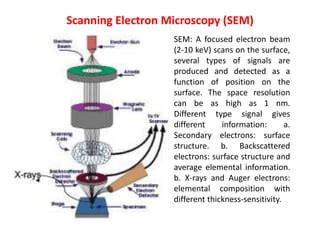
Oct 7, 2020 • Download as PPTX, PDF •. 4 likes • 787 views. JSPM Charak College of Pharmacy and Research. Follow. Transmission Electron Microscope (TEM), RESOLVING POWER, Scanning Electron Microscope, PRINCIPLE AND WORKING OF SEM, SEM SAMPLE PREPARATION, Limitations of Scanning Electron Microscopy (SEM), .Scanning electron microscopy Scanning Electron Microscopy - SEM. Sep 5, 2017 • Download as PPTX, PDF •. 16 likes • 3,777 views. Stephen Raj D. Scanning Electron Microscopy - SEM. Engineering. 1 of 68. Download now. Scanning Electron Microscopy - SEM - Download as a PDF or view online for free. 57. BIOLOGICAL APPLICATIONS OF SEM • Virology - for investigations of virus structure • Cryo-electron microscopy – Images can be made of the surface of frozen materials. • 3D tissue imaging - – Helps to know how cells are organized in a 3D network – Their organization determines how cells can interact. • Forensics - SEM reveals the . 32 likes • 4,733 views. P. piyush tripathi. Today, scanning electron microscopy (SEM) is a versatile technique used in many industrial labs, as well as for research and development. Due to its high lateral resolution, its great depth of focus and its facility for X-ray microanalysis, SEM is ofen used in materials science – including polymer . 5. Scanning Electron Microscopy (SEM) SEM: A focused electron beam (2-10 keV) scans on the surface, several types of signals are produced and detected as a function of position on the surface. The space resolution can be as high as 1 nm. Different type signal gives different information: a. Secondary electrons: surface structure.

34. THE SCANNING ELECTRON MICROSCOPE • To directly visualise the surface topography of solid unsectioned specimens. • Probe scans the specimen in square raster pattern. • The first scanning electron microscope (SEM) debuted in 1938 ( Von Ardenne) with the first commercial instruments around 1965.
SEM (SCANNING ELECTRON MICROSCOPE) SEM (SCANNING ELECTRON MICROSCOPE). 20823856 Özgen Buğdaycı 20824336 Elif Topçuoğlu 20823985 Yavuz Duran. Hacettepe University 12.04.2012. OUTLINE. Definiton of scanning electron microscope History Usage Area Instrumentation Sample preparation Working .
NAVI Javelins CS2 Female More about the team . Dota 2 Junior More about the team gotthejuice Taras Linnikov 19 years Carry .
scanning electron microscopy slideshare|Scanning electron microscopy