what is 2d echocardiogram with doppler|The 2D Echo Exam: What To Expect During Your Heart Ultrasound : Baguio The amount of information that’s shown on an echocardiogram is huge, and the answer to this question can be very long. To give a quick answer, an echocardiogram will show the overall function of your heart. What does this mean? Tingnan ang higit pa Find contact information, products, services, photos, videos, branches, events, promos, jobs and maps for Russ Marketing, Incorporated in Megacenter, Melencio Street .
PH0 · Two
PH1 · The 2D Echo Exam: What To Expect During Your Heart Ultrasound
PH2 · The 2D Echo Exam: What To Expect During Your Heart
PH3 · Echocardiogram: What It Shows, Purpose, Types, and Results
PH4 · Echocardiogram: What It Shows, Purpose, Types, and Results
PH5 · Echocardiogram: Types and What They Show
PH6 · Echocardiogram
PH7 · Doppler echocardiography
PH8 · Different Types of Echocardiogram
PH9 · 2D Echocardiogram w/ Doppler
PH10 · 2D ECHO/DOPPLER STUDY
ano sa english ang sagot sa.how will you aply it in real life situation. Last Update: 2023-07-11 Usage Frequency: . pagsagot ng pabalang sa magulang. English. answering parental rebuttal. Last Update: .
what is 2d echocardiogram with doppler*******An echocardiogram, or 2D echo or heart ultrasound an ultrasound examination that uses very high frequency sound waves to make real time pictures and video of your heart. Things that will be seen during a 2D echo test are the heart’s chambers, heart valves, walls and large blood vessels that are . Tingnan ang higit pa
The amount of information that’s shown on an echocardiogram is huge, and the answer to this question can be very long. To give a quick answer, an echocardiogram will show the overall function of your heart. What does this mean? Tingnan ang higit paI hope this guide has been able to answer some of the questions you might have had regarding what and echocardiogram is and what it does. If you still have any questions, please don’t hesitate to leave a comment below.or shoot me an email. I always . Tingnan ang higit pa

Generally speaking, no. A heart ultrasound does not show clogged arteries. The reason a 2D echo doesn’t show clogged arteries is because the coronary arteries are simply just too small to see with ultrasound. However, the larger arteries surrounding . Tingnan ang higit pawhat is 2d echocardiogram with dopplerGenerally speaking, no. A heart ultrasound does not show clogged arteries. The reason a 2D echo doesn’t show clogged arteries is because the coronary arteries are simply just too small to see with ultrasound. However, the larger arteries surrounding . Tingnan ang higit pa

Two-dimensional (2D) or three-dimensional (3D) echocardiogram. A 2D echo is the standard test, which shows your doctor images of your heart's walls, . Two-dimensional (2D) or three-dimensional (3D) echocardiogram. These images provide pictures of the heart walls and valves and of the large vessels connected .
Providers often combine echo with Doppler ultrasound and color Doppler techniques to evaluate blood flow across your heart’s valves. Echocardiography uses .Doppler echocardiography is a procedure that uses Doppler ultrasonography to examine the heart. An echocardiogram uses high frequency sound waves to create an image of the heart while the use of Doppler technology allows determination of the speed and direction of blood flow by utilizing the Doppler effect. An echocardiogram can, within certain limits, produce accurate assessment of the direction of bl.
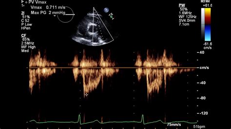
Doppler echocardiography. This Doppler technique is used to measure and assess the flow of blood through the heart's chambers and valves. The amount of blood .Doppler echocardiogram: This technique is used to measure and assess the flow of blood through the heart's chambers and valves. The amount of blood pumped out .2D Echocardiography/Doppler Study (Trans thoracic 2D Echo/Doppler Study) This is a non-invasive, painless and risk-free heart scan using high frequency ultrasound waves reflecting off various structures of the .
One of the many diagnostic tools available to Dr. Diego is Echocardiography (or ‘echo’, for short) with Doppler. This technology uses ultrasound to give the doctor a moving picture .Echocardiography in 2D. Two-dimensional (2D) ultrasound is the most commonly used modality in echocardiography. The two dimensions presented are width (x axis) and depth (y axis). The standard .Two-dimensional (2D) ultrasound is the most commonly used modality in echocardiography. The two dimensions presented are width (x axis) and depth (y axis). The standard ultrasound transducer for 2D .
what is 2d echocardiogram with doppler The 2D Echo Exam: What To Expect During Your Heart UltrasoundDoppler echocardiogram: This technique is used to measure and assess the flow of blood through the heart's chambers and valves. The amount of blood pumped out with each beat is an indication of the heart's functioning. Also, Doppler can detect abnormal blood flow within the heart, which can indicate a problem with one or more of the heart's .A Doppler echocardiogram uses a probe to record blood flowing through the heart. This technique enables Dr. Diego to see the heart and blood in motion. It can be used to evaluate the heart for many different disorders, including: Atrial fibrillation (irregular heartbeat) Damage to the heart muscle, possibly after a heart attack. An echocardiogram with strain is an ultrasound test that takes images of your heart and evaluates the function of your heart muscle (myocardium). Using this newer technique that measures heart muscle length during contraction and relaxation, a healthcare provider can identify subtle changes in your heart function. The 2D echo is a great way for doctors to identify heart structure problems. This video is an excellent opportunity to learn more about the 2d echo and see w.
Echocardiography is the use of ultrasound to evaluate the structural components of the heart in a minimally invasive strategy. Although, prior to the invention of today's routinely used 2-dimensional echocardiography, there was motion-based (M-mode) echocardiography. In 1953, Inge Edler, regarded as the father of .2D echocardiography, popularly called 2D echo, is a non-invasive test used to analyze the functioning and assess the sections of your heart. This test gives images of the different parts of the heart with the help of sound vibrations.It assists in checking damages, blockages, and blood flow rate. Doctors recommend regular 2D echo tests to .
Schneeballsysteme erleben eine Renaissance. DCPTG (.com) ist ein aktuelles Beispiel für solche betrügerischen Anlageversprechen.
what is 2d echocardiogram with doppler|The 2D Echo Exam: What To Expect During Your Heart Ultrasound