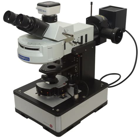
scanning lens microscope|1.4: Microscopy : Clark In laser-scanning microscopy often an off-the-shelf achromatic doublet is used as a scan lens which can reduce the available diffraction-limited field-of-view . Why us? 888casino is a multiple award-winning online casino that first opened its doors back in 1997. Today it boasts over 2000 top games enjoyed by over 17 million players worldwide. 888casino is licensed and regulated by the UK Gambling Commission and the Governments of Gibraltar and Malta. 888 Holdings plc has been listed on the London .
PH0 · Types of Objective Lens & Their Functions
PH1 · Types of Objective Lens & Their Functions
PH2 · Scanning electron microscope
PH3 · Scan Lenses for Laser Scanning Microscopy
PH4 · Optimal lens design and use in laser
PH5 · Microscopy: Intro to microscopes & how they work (article)
PH6 · Microscope Objective, Tube, and Scan Lens Tutorials
PH7 · Laser Scanning Microscopy Tutorial
PH8 · Introduction to Microscopes and Objective Lenses
PH9 · Fundamentals of Scanning Electron Microscopy in Life Science
PH10 · 3.1: Introduction to the Microscope
PH11 · 1.4: Microscopy
Vue.js 3.3 recently received the defineModels enhancement for two-way binding under the name defineModel, which has the exact same functionality.. You will find more information in our blog post: New in Vue.js 3.3: Two-Way Binding With defineModel Macro.However, opting for the defineModel functionality in Vue.js 3.3, as it is still an .The results shared here are extracted from the official sources of the Juwai Teer game. If you are searching for info about this game then you have landed on the right webpage because on this webpage you will get the all-important information related to Juwai Teer Result Today, Juwai Teer Previous Result List with Juwai Teer Common Number.
scanning lens microscope*******Types of Objective Lenses. Scanning Objective Lens (4x) Low Power Objective (10x) High Power Objective Lens (40x) Oil Immersion Objective (100x) What is the Difference between Ocular and Objective . The CLS-SL, SL50-CLS2, SL50-2P2, and SL50-3P lenses were optimized for use in Thorlabs' confocal laser scanning and multiphoton microscopy systems, and . Your microscope has 4 objective lenses: Scanning (4x), Low (10x), High (40x), and Oil Immersion (100x). In this lab, you will not use the oil immersion lens; it is .
In laser-scanning microscopy often an off-the-shelf achromatic doublet is used as a scan lens which can reduce the available diffraction-limited field-of-view . Learn how to use the microscope to view slides of several different cell types, including the use of the oil immersion lens to view bacterial cells. Early .A microscope is a special optical device designed to magnify the image of an object. Depending on the type of microscope, it may project the image either onto a human eye or onto a recording or video device. As an . Scan Lenses for Laser Scanning Microscopy. Telecentric Scanning Lenses with Flat Image Planes. Minimal Optical Aberrations and Low F-Theta Distortion. Large Maximum Fields of View up to 28.9 x 28.9 .Introduction. The scanning electron microscope (SEM) is a powerful tool that uses high energy electrons, instead of visible light, to methodically scan the surface of a specimen . Laser scanning microscopy (LSM) is an indispensable imaging tool in the biological sciences. In this tutorial, we will be discussing confocal fluorescence imaging, multiphoton excitation fluorescence .But most commonly, when talking about types of objective lenses we are referring to the different magnifications and purposes of the four most common types of microscope objective lenses on compound light .
Every microscope has an eyepiece lens, which is the lens at the top that you look through. A tube connects the eyepiece lens to objective lenses, which enhance the magnification power of the eyepiece lens. . A .imaging. The lens system consists of condenser and objective lenses, apertures and scanning coils. Optical focusing in light microscopes occurs with a glass lens when the air-glass interface causes refraction of light rays and the spherical shape of the lens leads to focusing. In an electromagnetic lens, however, current1.4: Microscopy scanning electron microscope (SEM), type of electron microscope, designed for directly studying the surfaces of solid objects, that utilizes a beam of focused electrons of relatively low energy as an electron probe that is scanned in a regular manner over the specimen. The electron source and electromagnetic lenses that generate and .scanning lens microscope 1.4: Microscopy The most familiar type of microscope is the optical, or light, microscope, in which glass lenses are used to form the image. Optical microscopes can be simple, consisting of a single lens, or compound, consisting of several optical components in line. The hand magnifying glass can magnify about 3 to 20×. Single-lensed simple .A scanning probe microscope does not use light or electrons, but rather very sharp probes that are passed over the surface of the specimen and interact with it directly. This produces information that can be assembled into images with magnifications up to 100,000,000⨯. . on a microscope, the lens closest to the eye (also called an eyepiece .
In the simplest beam-scanning confocal configuration, a scan mirror is located in the rear focal plane of a scan lens, which is conjugate with the rear focal plane of the objective. Illustrated in Figure 3(a) is a single-mirror arrangement, which includes the tube lens required by an infinity-corrected objective.
Magnification is a measure of how much larger a microscope (or set of lenses within a microscope) causes an object to appear. For instance, the light microscopes typically used in high schools and colleges magnify up to about 400 times actual size. So, something that was 1 mm wide in real life would be 400 mm wide in the microscope image.Scanning electron microscopy is a characterization technique that images and analyzes a specimen by scanning an accelerated electron beam, followed by selectively collecting and recording secondary electrons, back-scattered electrons, and other signals arising from the beam and specimen interactions. A modern scanning electron microscope (SEM .
Microscopes often contain multiple objectives on a rotatable nosepiece, for example: a scanning lens with only 4 × magnification, an intermediate 10 × small objective lens, and; a high-resolution large objective with 40 × or 100 × magnification. The eye piece may be fixed or also exchangeable.
On a microscope, there are usually 3 – 4 objective lenses. These lenses sit on a rotating turret just above the microscope’s stage. The objective lens band colors are universal in order to standardize the function of a .
Near-field scanning optical microscopy is classified among a much broader instrumental group referred to generally as scanning probe microscopes ( SPMs ). All SPMs owe their existence to the .

The RP Photonics Buyer's Guide contains 26 suppliers for scanning lenses.Among them: EKSMA OPTICS. Our F–theta scanning lenses are designed for laser marking applications with Nd:YAG laser’s .
Abstract. When used appropriately, a confocal fluorescence microscope is an excellent tool for making quantitative measurements in cells and tissues. The confocal microscope’s ability to block .

Microscope Objectives or Objective lenses are in many ways the heart of the microscope, and are typically mounted on a rotating nosepiece or turret to enable easy selection. Many microscopes will be equipped with a scanning objective (4x), a low power objective (10x), a high power objective (40x), and perhaps even an oil immersion objective lens.
Laser scanning microscopes use laser illumination to generate high-resolution, high-contrast 3D imagery of samples by scanning them point by point. Two common types of laser scanning microscopes include confocal laser scanning microscopes and multiphoton laser scanning microscopes. While both use lasers to excite the sample, . In this section we will examine microscopes, instruments for enlarging the detail that we cannot see with the unaided eye. The microscope is a multiple-element system having more than a single lens or mirror (Figure 26.4.1 26.4. 1 ). A microscope can be made from two convex lenses.
Make sure to dry off the slides and coverslips with paper towels (not the expensive lens paper) before preparing your wet mount slides. Get a dry microscope slide and cover slip. Put a drop of methylene blue on the slide. Gently scrape the inside of your cheek with a toothpick and swirl it in the dye on the slide.
Free bonuses from the best online casinos. At Vegas Expert, I focus on selecting the very best online casino options and helping even the most demanding players find one that meets all of their expectations.. That is why this page only contains no deposit bonuses offered by tested online casinos with a good reputation and fair approach to gambing. .
scanning lens microscope|1.4: Microscopy